
In veterinary medicine radiographs are used regularly to diagnose and monitor the progression of many medical conditions. At the Victoria Veterinary Clinic radiography is also used as a surgical tool. During dental and orthopedic surgeries x-rays are taken at various intervals to help guide the surgeon, in the case of dental surgery, x-rays can help determine which teeth need to be removed as well as areas where bone loss is present. During an orthopedic surgery it can be used to help determine the correct position of a bone or pin which allows for a more precise result.
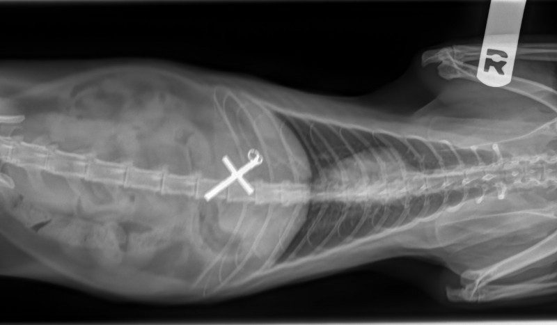
 Dog with bladder stones
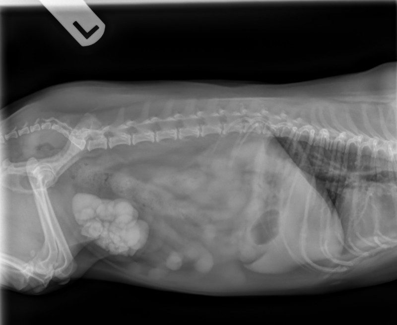
Foreign Body in a cat
X-ray technology was first discovered by German physicist Wilhelm Röntgen in November of 1895. His work included experimentation with electronic beams. During one experiment he noticed a fluorescent screen in his laboratory had begun to glow. When the rays were blocked with lead he found that he was able to see the bones of his hands. He published the results of these experiments six weeks later along with a photograph of this phenomenon, which showed the bones of his wife’s hand. He named his discovery “X-ray”.
Once these findings were made public, it took no time at all to create x-ray machines. X-ray machines send individual x-ray particles through the body, and these images are recorded on film, or more recently on computer systems. Dense structures, such as bone, block most of the particles and they appear white.
In 1896, not long after Wilhelm Röntgen’s discovery, three college students in North Carolina convinced a janitor to let them into a physics laboratory where they took x-rays of various objects, resulting in the first x-ray image in North America. They took images of a pin, two bullets and two rings inside of a pill box, a cadaver finger stuck with pins and wearing a ring, a hollowed out egg with a button inside and a magnifying glass.
The exact same day on the other side of the world John Hall-Edwards was the first to use x-rays under clinical conditions when he radiographed a needle stuck in the hand of a co-worker prior to its removal.
Scientists and physicians were quick to realize the benefits of x-rays, but not on the harmful effects of radiation. Within a few years, reports of burns, skin damage and cancer after exposure to x-rays emerged. Today safer methods are used, including specialized training and attire for professionals, using the smallest quantity of radiation needed to reduce exposure, and modern x-ray machines that only emit radiation while it takes a picture.


